Sports injury management

Sports injury management

When it comes to sports injury management, most people are familiar with the old acronym RICE (rest, ice, compression, elevation) for soft tissue injury management, but what is the evidence for it, and can we do better?

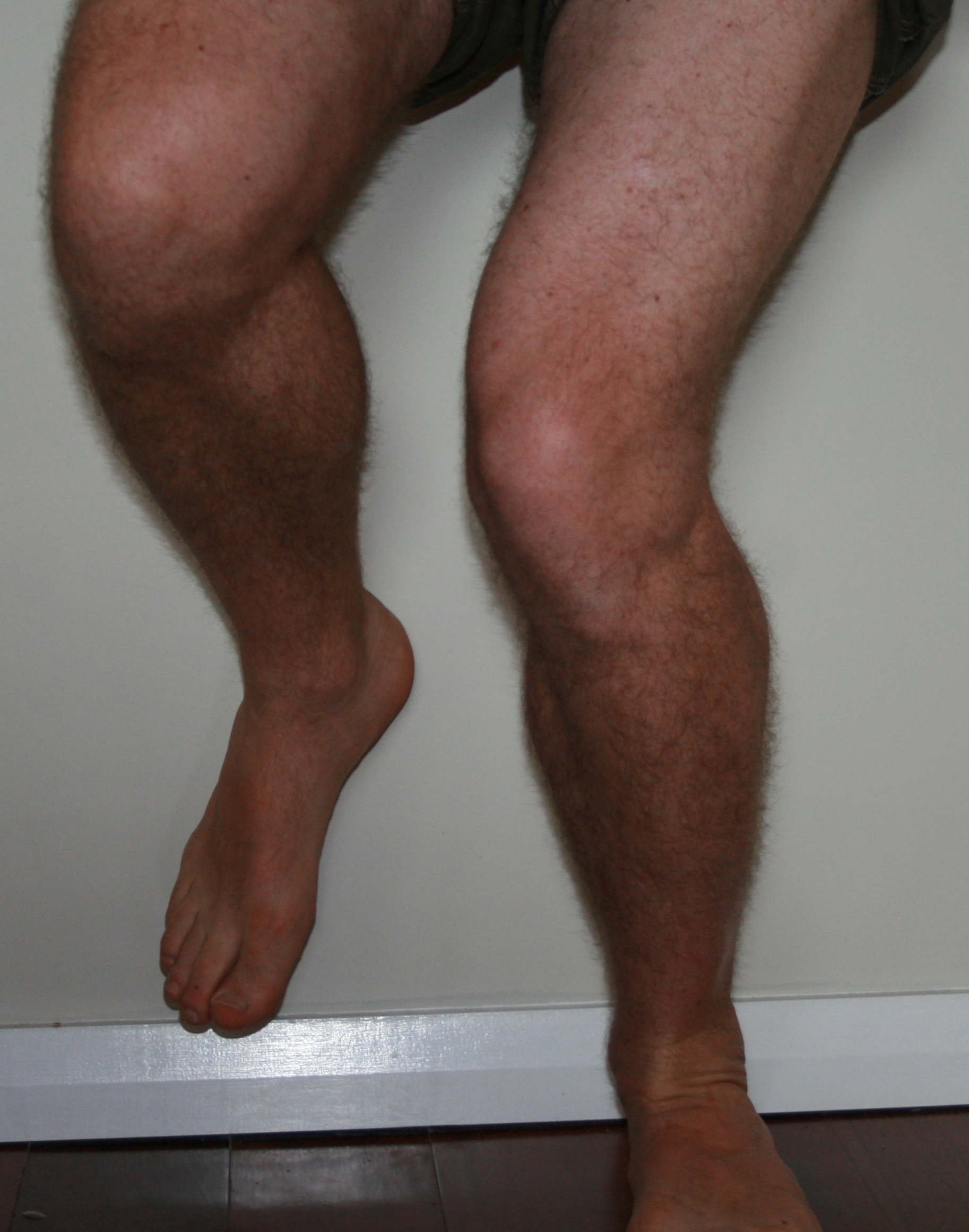
Complete rest is rarely necessary following simple soft tissue injuries. For example, following a simple ankle sprain, a person who continues to move the ankle and weight bear as tolerated will have accelerated recovery compared to someone who avoids weight bearing and keeps the ankle still.

Ice is often used immediately following a soft tissue injury. Ice does not reduce swelling. If applied correctly, ice can reduce the temperature of surrounding tissues enough to reduce the nerve’s ability to carry information back to the brain, thereby reducing pain. Unfortunately, ice can interrupt the natural inflammatory response following an injury and possibly delay the healing response. The consensus is: only ice following an injury if the pain is intolerable – then ice it properly by making a bag of crushed ice and water.

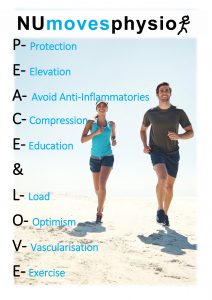
The new recommended acronym for soft tissue injury management. PEACE (Protect, Elevate, Avoid anti-inflammatories, Compress and Educate) refers to the steps immediately following injury. In the next few days, the injury needs a bit of LOVE (Load, Optimism, Vascularisation and Exercise). For more information on how to best manage your injury, contact us on 49216879 or admin@numoves.com.au

For the full article visit the BJSM editorial: https://bjsm.bmj.com/content/54/2/72

Thoracic pain

thoracic pain

The thoracic spine is your mid back between lower back and neck. Each of the 12 vertebrae of your thoracic spine has a rib on each side. Acute thoracic spine pain can be very debilitating. When it hurts to breathe the joints between the ribs and the thoracic vertebral body and transverse process can be the source. Postural loading and stress are common causes of this type of pain. Another is trauma in sport, but you also need to make sure there are no rib or other fractures in these cases.

Simple analgesics are usually advised at first. Sometimes taping can limit the pain by restricting painful movement when pain is bad but shouldn’t be used for more than a few days. The next step is to get moving via walking in water or on land followed by stretches. At this stage massage and joint mobilisations can improve your overall movement and increase the speed of your recovery.

Get good advice early is the quickest way to get back to normal once you have acute thoracic pain. This also includes analysis of the causes and implementing movement-based prevention strategies. Read more here on how we can help or contact us on 4921 6879.

Knee pain – what is valgus collapse

One of the most common joint problems associated with exercise is pain in the front of the knee (patella). It is commonly labelled runners knee or jumper’s knee but often just walking on slopes or stairs can be the source of the pain. If we exclude degeneration of the cartilage under the patella (which is diagnosed with MRI scans), this type of knee pain can be effectively managed.

Knee pain and valgus collapseThe greatest external load on the front of the knee is due to poor movement pattern, where the knee goes inwards towards the other leg rather than straight ahead in line with the middle of the foot. This is termed valgus collapse.

Valgus collapse is a strength and movement problem which can be corrected with specific exercises targeting the right muscles. A thorough physiotherapy assessment should consider:
• Muscle length (especially the outer thigh)
• Muscle strength (particularly at the hip)
• Foot mechanics / posture
• And the way you move (muscle control or coordination)

The treatment is a series of pain free exercises that address the cause of the problem. Taping the knee is a short term option for reducing pain. Sometimes changes in footwear and / or orthotics can also assist with pain reduction, but neither replaces getting stronger and moving better. If you have persistent pain in the front of the knee, have it assessed and get a prescribed exercise plan.

Common training mistakes – how to avoid injury

running and physiotherapy

Exercise and sports related injuries are common. Some sports such as rugby come with higher risks and frequency of injury. General exercise often has low risk of injury but that depends on how hard we push ourselves and the type of exercise we do. It doesn’t matter whether it is a team sport, an exercise class, a personal training session or an individual session, an injury will limit your participation for a period of time.

One common source of injury is ‘training error’. This is usually doing too much, too soon but can also be related to technique (e.g. squat, running) or equipment. Technique is the way you move relative to the load or complexity of the movement.

There is a fine balance between being challenged sufficiently to enjoy exercise and avoiding ‘training error’. A simple message is the more you know your body at each stage of life the less likely injury will occur. This means train at a level of load suited to your body; gradually increase the level of exercise over several weeks; listen and adjust to pain especially if it is in a joint or the spine; and use mirrors to watch how you perform a gym exercise to ensure the technique is correct. It’s simple advice but it works if you want to reduce the chance of injury.

Stress fractures in runners – 3 minutes of what, why & when

female runner


What is a stress fracture: Normal bone responses to repetitive stress can be divided into: normal response, stress reaction, and stress fracture.

Normal response: Osteoclasts are bone cells that remove bone tissue known as ‘bone resorption’. Whilst osteoblasts are cells that create ‘bone formation’. Bone is a dynamic tissue that is constantly being reshaped by osteoblasts and osteoclasts working in balance.

Stress Reaction: Repeated bone stress without appropriate rest causes osteoclastic activity to outstrip osteoblastic activity.  This osteoclast / osteoblast imbalance initially results in microfractures which when investigated shows bone marrow oedema.
 
Stress fracture: A stress fracture is when the repeated stress and imbalance continues and the microfracture progresses into a true break in the cortical bone.
 
How are they diagnosed: A clinical examination of factors such as training loads, biomechanics, location of pain, and bony tenderness, will indicate whether a bony stress reaction / fracture is a possibility. Following this various imaging options are MRI (which can show bone marrow oedema in stress reaction stages and microfractures once stress fracture develops); CT scans (not as sensitive as MRI and have associated radiation as per an XRay); Xray (which frequently doesn’t show up stress fractures in the early stages – up to 3 weeks); and bone scans (very good at detecting stress fractures but are time consuming, nonspecific and are a poor choice to monitor recovery).
 
Who is most at risk: The female athlete triad is a combination of low bone density, nutritional issues, and menstrual irregularities. Together these represent the highest risk of developing stress fractures in the female runner. However any one of these alone can also lead to a stress fracture when combined with running.
 
What else increases your risk:

  • Rapid increase in running distances
  • Lack of rest / recovery time during a period of running training
  • Inappropriate footwear relative to the foot type of the individual
  • Nicotine smoking

What to look out for:

  • Pain during or after running / exercise
  • Bony tenderness
  • Nutrition – how balanced is your diet
  • Training – running distances per week and speed of increases
  • Bone density – do you know yours? (especially if you are female and run a lot)
  • Shoes – despite the debates surrounding ‘support’ vs ‘free’ shoes, the right type of footwear is important for a lot of people!

Management: If you have pain with running organise an assessment as soon as possible. The best management is prevention but if they occur the treatment depends on which bone it is. Some stress fractures have greater risks associated and need orthopaedic specialists involved, whilst others are lower risk and can be managed conservatively. The most common strategy is to immobilise the area and often to remove any weight bearing stress through the bone.

McCormick et al (2012), Stress fractures in runners. Clin Sports Med, 31.

Do you know how to correct your posture?

correct postureMaster the simple things and you will get the best outcomes!
We all have our own unique postures – some with increased curves, others with reduced curves, and sometimes with sideways curves (scoliosis). Posture should also be considered relative to whether you are sitting, standing or dynamically moving. Achieving a biomechanically correct spinal posture is important for several reasons.

Firstly, in helping our clients we often find that achieving the correct posture can reduce pain and discomfort associated with sustained standing and sitting postures. For those who have to stand for long periods in retail or hospitality, an increased curve in the lower back can lead to pain. A simple correction is to lean up against the wall slide down to bend the knees slightly and flatten the lower back to the wall. If this eases the discomfort we need to focus on what creates this posture type – tight hip flexors, poor tone or weakness in the lower abdominals and gluteal (buttock) muscles.

Alternatively those who get discomfort in the lower back, mid back or neck with sitting at the computer, it often is associated with the ‘lazy, slouched posture’ where the curve in your lower back (normally known as a lordosis) is reversed and there is one arched curve (instead of two) from base of the spine to base of the neck. The head then protrudes forwards and the shoulders become rounded. Again a simple correction is pelvic tilting – rolling the pelvis forwards over your hip bones. Getting that lordosis curve back in the lower back will often reduce pain and allows for the mid back (thoracic spine) and neck posture to be corrected. You have to get a stable base to work from and slouched is not a stable base.

Secondly correct posture increases the efficiency and safety of how we move, for example with lifting. The squat lift (knees fully bent) or semi-squat lift (knees bent to 90 degrees) are both acceptable methods of lifting. If you keep your lower back straight and bend at the hips and knees it will engage the gluteal muscles and provide stability to the pelvis and lower back.

The big message, it is OK to relax your posture at times so long as it isn’t occasions such as when you are lifting loads or sitting for prolonged periods.


Shoulder Impingement and the Rotator Cuff

xray imageWhat is the rotator cuff?
The rotator cuff muscles consist of 4 small muscles that work together to keep the shoulder joint centred – supraspinatus (most commonly associated with a rotator cuff tear); infraspinatus and teres minor (the external rotators); and subscapularis (the internal rotator and least understood but arguably the most important).

Why is it important?
Imagine the shoulder joint as a golf ball sitting on a golf tee. It requires the small rotator cuff muscles closest to the joint to stabilise it so the larger muscles can move the arm efficiently.

What is shoulder impingement?
Impingement is the most common pathology seen in the shoulder, which involves the tendons and other soft tissues get squished under the acromion (a bony arch forming a roof above the shoulder joint).

How do we manage it?
The management largely depends on the cause. Often a muscle imbalance is created by repetitive use of the arm over shoulder height (throwers or swimming). The muscle imbalance can be associated with the scapula (resulting in scapula winging or lack of rotation control) which requires specific exercise to stabilise the shoulder blade region.

Additionally a muscle imbalance of the rotator cuff can be created by the pain associated with impingement symptoms. This can be identified via a physical assessment of active movement control versus passive range available and if clinically relevant to the symptoms will result in improvement within a 2 week period. Occasionally the acromion is shaped in a hook fashion which increases the chance of impingement and this can require surgery (acromioplasty) to shave away and make more space in the area.

The concept that muscle imbalance and fatigue can lead to shoulder impingement has been validated over the years, and supports the use of rotator cuff strengthening exercises as an effective treatment for shoulder impingement.


Eccentric exercise for the achilles tendon

calf muscle and achilles tendonExercise for the achilles tendon is most commonly done via eccentric exercise which involves contracting a muscle as it lengthens. The opposite is termed a concentric contraction where the muscle shortens as it contracts. The force generated during an eccentric exercise is higher than a concentric contraction. The subsequent loading of a tendon occurring with an eccentric contraction is the basis for the Alfredson protocol of Achilles tendon rehabilitation.

An example of an eccentric contraction is for the calf muscle and achilles tendon complex. If you stand with both forefeet on a step and push up onto your toes, a concentric contraction results where the muscles shorten as they contract. If you then lift one foot off the ground and lower yourself down on the other foot, an eccentric contraction occurs with the muscle contracting but lengthening at the same time. Higher force is generated in the Achilles tendon during this movement which is the basis for the Alfredson protocol. Repeated loading of the tendon via this exercise results in the tendon being able to tolerate higher loads. Not all achilles tendon problems are suited to this approach. A good place to start is an individual assessment by a NU Moves physiotherapist. 

Don’t Be Limited By Your Tendons: Achilles Tendonitis

achilles tendon physio newcastle

Achilles tendonitis … if you have suffered from it you know how much it can limit you with everyday life activity. The actual diagnosis is now termed ‘tendinopathy’ rather than tendonitis as recent evidence has found there is not an actual inflammatory process within the tendon. Regardless of the name, it refers to pain located usually up to 5cm up from the back of the heel. There are several different types of Achilles tendinopathy so if you suspect you have this problem it is a good idea to get a physiotherapist to confirm the diagnosis and type of presenting problem.

The good news is simple inexpensive treatment is available and quite effective in getting you active again. We commonly recommend 3 strategies that help:

  1. Despite the lack of bleeding or inflammatory cells present in a tendinopathy, applying ice safely to the local area still does provide benefit to many people. Simple things can be beneficial even if the understanding of why isn’t so simple. Put simply the benefit of ice is to reduce pain which assists beneficial exercise to be undertaken.
  2. Relative rest is also advised when managing tendon pain. This doesn’t mean complete rest from the aggravating activity but usually a reduction in the task that aggravates the pain.
  3. Active exercise based rehabilitation is the key to regaining strength and reducing pain with everyday life and exercise. This involves gentle and progressive loading exercises for your Achilles’ tendon known as the Alfredson protocol. The exercise is a type of eccentric exercise that is optimally performed with knees straight and knees bent and involves 3 sets of 15 repetitions of each. There may be soreness or pain that can last for a day, but the soreness should reduce as you progress the exercises over the course of weeks. Working with your physio will guide how much soreness is OK and when the protocol should be modified.
  4. To compliment these 3 treatments we also provide stability exercises for the core and legs muscles to compliment the Achilles exercises. For example, a lack of strength at the hip can increase the requirements and load on the calf and Achilles region. A more comprehensive and effective program will find any such imbalances and help if the goal is regular or more intensive exercise. To learn more, read our What is Eccentric Exercise? article.

If you have an achilles problem, start with a physio assessment and ask if the Alfredson protocol is appropriate for you.

Low back pain – Is it in the way you move?

woman doing fitnessMedical literature places all those with low back pain that have no pain down the legs or other nerve or medical features into one group “non-specific low back pain”. It is a large group (approx 80%) of all people who have low back pain. So how do we best help such a large group of different people with non-specific low back pain? Every person is different in their physical makeup and past experiences.  We cannot separate the ‘who’ you are from the ‘what’ is wrong when answering this question.

 5 areas worth considering when helping someone with non-specific low back pain.

1. How much sitting do you do? As a nation, we are now sitting more than ever before thanks mostly to computers. Remember the message from Dr Mike Evans and his 23 1/2 hrs video! If you haven’t watched it, click here to learn what’s the single best thing we can do for our health.

2. How much movement/exercise do you do? Too much of any position can make us sore, and simply doing something different can offset that. Walking to counteract the sitting can help those who have pain with sitting. If that is you, start a regular walking program.

3. The way you move can predispose you to pain. Dr Peter O’Sullivan is a world leader in researching back pain and remains a practicing physiotherapist in WA. His research looks into sub-classifying mechanical low back pain into which movements increase it and improving how you move to reduce it.

4. Core strength – this actually has the least amount of research behind its benefits. Sometimes strengthening the back and abdominal muscles does work (as evidenced by those doing pilates) but other times it creates pain.

5. Stress levels – this is a common contributor to low back pain. Occasionally it is the main cause of the pain but often being stressed just makes your pain worse. There are lots of resources to help in this area but starting with your GP is a good option.

Management needs to identify and address all of these factors. Get moving if you aren’t already (if the pain allows you). Get out of the chair as much as you can. Look at what movements or postures make the pain worse – that’s where your physio can help the most! Consider your stress levels relative to the pain. Lastly you can start core exercise/pilates if there is no increase in pain – note there are many types of core exercises and choosing the right level and having the correct technique is important. Sometimes it helps because it’s just another method of getting you out of the chair and active.

It’s a lot to consider but if you have back pain that is affecting your life then you need a physiotherapy assessment. We will get you moving in the right way. Let us know if we can help at 4921 6879 or email admin@numovesphysiotherapy.com.au.